
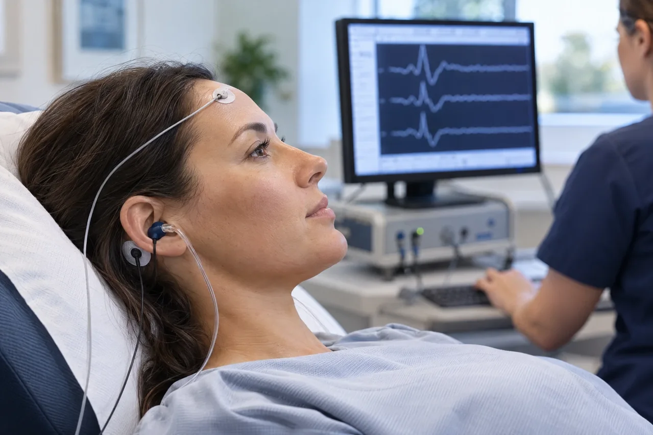
The study & its electrophysiologic principle
Brief clicks or frequency-specific tones are delivered separately to each ear through insert earphones or other transducers. Surface electrodes at the scalp and ear or mastoid detect small voltage changes; repeated sweeps are averaged to distinguish the time-locked response from background activity.
The short-latency waveform reflects synchronized auditory-nerve and ascending brainstem activity following sound stimulation. Waves are conventionally labeled I through V, but peaks can have multiple contributing neural generators rather than a simple one-structure/one-wave identity. Interpretation considers the complete waveform and recording conditions.
Common clinical applications
Qualified clinicians may use ABR to estimate hearing sensitivity or support auditory assessment in patients who cannot provide dependable behavioral responses. Frequency-specific responses can complement audiologic testing; a screening result is not a diagnostic hearing evaluation.
In appropriate contexts, ABR may contribute evidence about auditory-nerve or brainstem dysfunction and selected intraoperative monitoring. Findings can be affected by conductive or cochlear hearing loss and should be integrated with history, examination, audiometry, otologic assessment, and other studies.
What you may expect
You will usually recline while small surface electrodes and earphones are placed. Brief clicks or tones are recorded while you relax, keep still, or sleep. Placement may cause temporary skin pressure or mild adhesive discomfort; sensation varies, and pain-free testing cannot be guaranteed.
General preparation may include clean hair and quiet rest. Do not stop, change, or skip prescribed or over-the-counter medicines unless the clinician managing them specifically tells you to. Sedation, when considered, has separate medical requirements and risks.
Time depends on the protocol and recording quality. Results may require review rather than immediate discussion, and the test cannot promise an outcome.
Explore the technical detail.
Open the sections below for anatomy, physiologic parameters, methodology and clinical limitations.
Anatomical & physiologic context
Auditory input originating in the cochlea activates the auditory nerve and then distributed pontine and midbrain pathways. ABR records the resulting synchronized neural activity; it does not directly measure cochlear hair-cell function. Wave I is chiefly associated with distal auditory-nerve activity, while later components reflect distributed auditory-nerve and brainstem generators.
ABR characterizes sound-evoked neural timing, not cerebral background activity or routine peripheral motor and sensory conduction. Stimulus delivery, hearing status and recording conditions are essential to understanding the response.
Important physiologic parameters
- Waveform presence, reproducibility, morphology, and polarity under the selected montage and stimulus condition.
- Absolute and interpeak latencies, interpreted with age, hearing status, temperature, technical factors, and validated laboratory norms.
- Response amplitude and amplitude relationships, recognizing that amplitudes are sensitive to electrode placement, impedance, noise, and physiologic variation.
- Stimulus type, frequency content, intensity, polarity, repetition rate, ear-specific presentation, and masking where indicated.
- Electrode montage, impedance and artifact control, replicate traces, noise level, and adequacy of averaging for reliable waveform identification.
General methodology
The patient reclines while electrodes are applied to prepared skin at scalp and ear or mastoid sites. Each ear is stimulated in turn, and the system averages responses. The operator checks electrode contact, artifact, stimulus delivery, and repeatability; protocols vary with the clinical question and age.
Data acquisition produces waveforms and measurements. Licensed medical interpretation, clinical correlation, diagnosis, and treatment decisions are separate responsibilities of the authorized healthcare professional reviewing the complete case. Apex NeuroMetrics informational content is not an independent diagnosis or treatment service.
Clinical relevance & limitations
ABR measures sound-evoked neural timing without requiring a button press or spoken response. A detectable or absent response is interpreted in relation to stimulus level, hearing status, technical quality, and norms; a waveform finding alone does not establish disease.
Recorded data complement symptoms, history, examination, audiologic findings, and other studies. Licensed medical interpretation is needed to determine significance and next steps; acquisition itself is not diagnosis or treatment.
- Limitations include hearing loss, middle-ear conditions, age, alertness, artifact, and normative variation. ABR is not a definitive diagnosis by itself and does not replace clinical assessment.
- ABR is not interchangeable with other electrophysiologic methods; test selection, hearing assessment and interpretation require an appropriate clinical framework.
Information, within appropriate scope.
Recorded physiologic data support broader clinical evaluation. Licensed medical interpretation, diagnosis and treatment are separate clinical responsibilities. Specific service availability, professional qualifications and arrangements must be verified.
Educational references
Selected sources support the educational discussion. They do not imply affiliation, endorsement or an individual clinical recommendation.
- Guideline 9C: Guidelines on Short-Latency Auditory Evoked Potentials, American Clinical Neurophysiology Society (opens in a new tab)
- BAER - brainstem auditory evoked response, UCSF Health (opens in a new tab)
- Brainstem Auditory Evoked Response or Auditory Brainstem Response (Unsedated), Children's Minnesota (opens in a new tab)
- Guidelines and Consensus Statements, American Clinical Neurophysiology Society (opens in a new tab)